Science
Transforming Histology: Advancements in 3D Imaging Technology
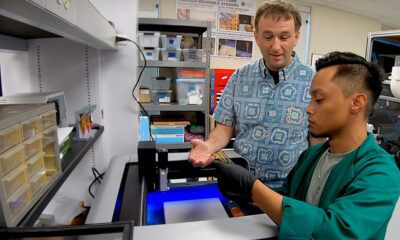
Advancements in histology are paving the way for enhanced medical diagnostics through the integration of 3D imaging technology. Traditionally, physicians have relied on microscopic tissue analysis, which involves cutting tissue into ultrathin sections and staining them with special dyes. This method allows doctors to determine whether tissue is pathologically altered and identify the presence and type of tumors, influencing therapeutic decisions.
Historically, the process of histological examination has been labor-intensive and time-consuming. The use of light microscopes has been pivotal in diagnosing conditions, particularly in assessing whether all altered tissue has been removed during surgical procedures. In recent years, however, innovations in imaging technology have begun to transform this landscape, offering a more efficient and accurate means of analysis.
Revolutionizing Diagnostic Processes
The introduction of 3D imaging technology into histology has the potential to revolutionize how pathologists conduct diagnostics. By creating detailed three-dimensional images of tissue samples, this technology allows for a more comprehensive view of the sample’s structure. This capability not only enhances the ability to identify tumors but also aids in understanding the tumor’s microenvironment and how it interacts with surrounding tissues.
Research indicates that 3D imaging can significantly improve diagnostic accuracy. For instance, a recent study published in the Journal of Histological Science highlighted that pathologists using 3D imaging techniques were able to diagnose certain types of tumors with a precision increase of up to 25% compared to traditional two-dimensional methods. This advancement is critical in an era where timely and accurate diagnosis can be a matter of life and death.
Moreover, the application of this technology extends beyond tumor identification. Surgeons can utilize 3D imaging to assess the extent of tissue alteration during operations, ensuring that all affected areas are addressed. This can lead to more successful surgical outcomes and reduce the likelihood of recurrence.
Implications for Future Medical Practice
The implications of integrating 3D imaging technology into histology are profound. As the healthcare industry increasingly embraces digital solutions, the shift from conventional histological methods to advanced imaging techniques could lead to significant improvements in patient care. Enhanced diagnostic capabilities may result in more personalized treatment plans, which are tailored to the specific characteristics of a patient’s tumor.
Furthermore, the adoption of 3D imaging could pave the way for advancements in research, enabling scientists to explore tissue characteristics in greater depth. This could lead to new insights into disease mechanisms and the development of targeted therapies.
In conclusion, the evolution of histology through 3D imaging technology represents a significant leap forward in medical diagnostics. As research continues to validate the benefits of these techniques, healthcare providers are likely to adopt these innovations more widely, ultimately improving patient outcomes. The transition from traditional methods to advanced imaging not only enhances diagnostic precision but also fosters a deeper understanding of complex tissue structures, marking a new era in the field of pathology.

Shark Bites Surge Linked to Climate Change and Ecosystem Shifts

NASCAR Clash Delayed Due to Snow, Rescheduled for Monday

Boeing and Lockheed Martin Battle for Fighter Jet Dominance

Golden Link Chapter No. 204 Honors Kijahfha Mahoney with Service Award

Gigachad Price Falls to $0.0025 Amid Market Fluctuations

Exploring Bortus: The Complex Alien of ‘The Orville’

Harry Potter Quiz Links Personality Traits to Entrepreneurship

Colorado Voters React to Trump’s Immigration Actions Amid Midterms

Donaldson Capital Management Boosts Stake in UnitedHealth Group

University of Hawaiʻi Joins $25.6M AI Project to Monitor Disasters
Foreign Inflows into Japan Stocks Surge to ¥1.34 Trillion

Hudson Williams Gains Popularity as Breakout Star on Heated Rivalry

Boeing’s Merger with McDonnell Douglas: A Strategic Move Explained

$1.25M Grant Advances Hawaiʻi’s Real-Time Hazard Monitoring

Sydney Sweeney Embraces Body Positivity Amid Hollywood Challenges

BOYNEXTDOOR’s Jaehyun Faces Backlash Amid BTS-TWICE Controversy

French Film Explores Group Therapy in ‘Group – The Schopenhauer Project’

Urgent Farewell: Joleen Chaney Leaves Legacy at KFOR
-
Science3 months ago
University of Hawaiʻi Joins $25.6M AI Project to Monitor Disasters
-
 Business3 months ago
Foreign Inflows into Japan Stocks Surge to ¥1.34 Trillion
-
 Entertainment2 months ago
Hudson Williams Gains Popularity as Breakout Star on Heated Rivalry
-
 World3 months ago
Boeing’s Merger with McDonnell Douglas: A Strategic Move Explained
-
 Science2 months ago
$1.25M Grant Advances Hawaiʻi’s Real-Time Hazard Monitoring
-
 Entertainment3 months ago
Sydney Sweeney Embraces Body Positivity Amid Hollywood Challenges
-
 Top Stories3 months ago
BOYNEXTDOOR’s Jaehyun Faces Backlash Amid BTS-TWICE Controversy
-
 World3 months ago
French Film Explores Group Therapy in ‘Group – The Schopenhauer Project’
-
 Top Stories3 months ago
Urgent Farewell: Joleen Chaney Leaves Legacy at KFOR
-

 Top Stories3 months ago
Marc Buoniconti’s Legacy: 40 Years Later, Lives Transformed
-

 Lifestyle4 months ago
Kelsea Ballerini Launches ‘Burn the Baggage’ Candle with Ranger Station
-

 Top Stories3 months ago
Carson Wentz Out for Season After Shoulder Surgery: Urgent Update